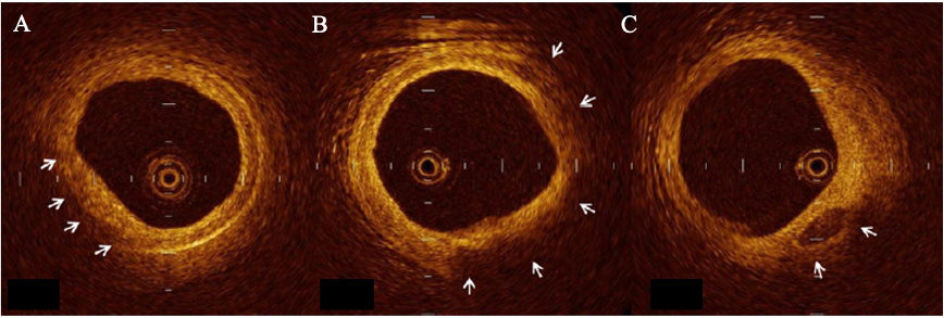
Intracoronary imaging by optical coherence tomography (OCT)
Although it is the exam of choice for the visualization of the coronary arteries, a coronary angiogram alone is unable to directly visualize atherosclerotic plaques within the arterial wall. For over a decade, new intracoronary imaging technologies have been developed to allow direct and detailed visualization of the arterial wall and atheroma plaque. Using infrared light, optical coherence tomography (OCT) enables visualization of the appearance of a plaque, characterization of its contents, and better understanding of the mechanisms responsible for the occurrence of a heart attack as well as validation of the optimal apposition/deployment of a stent.
How does OCT work?
OCT is often performed after the diagnostic coronary angiogram. The procedure is similar to a coronary angioplasty except that the balloon is replaced by a small infrared imaging catheter that is placed in the diseased coronary artery over a metal guidewire. This procedure is painless.

It is dangerous?
Complications related to intracoronary imaging are the same as that of the coronary angioplasty.
Rotational atherectomy – The RotablatorTM
Rotational atherectomy is a technique involving the ablation of an atheroma plaque using a rapidly rotating olive-shaped device with its distal end is covered with diamond micro fragments. This tool is used in 3-5% of coronary angioplasty procedures. It is useful in the presence of highly calcified lesions, stenoses that cannot be crossed with balloons or stents and for lesions that are resistant to high-pressure balloon inflation. The microparticles released into the bloodstream during tissue ablation (5 microns) are naturally removed from the body and are absorbed by the reticuloendothelial system and eliminated by the spleen.

How does rotational atherectomy takes place?
The use of rotational atherectomy does not change the course of coronary angioplasty. It is only an additional tool used during the procedure. The sound generated by the device will remind you of the tool used by the dentist. Rarely the use of this device will generate brief chest pain identical to that felt during your angina attacks.
Is it dangerous?
Complications related to the use of rotablator are the same as that of the coronary angioplasty. Complications specifically related to the use of this device are rare (0.2 to 0.6% of procedures).
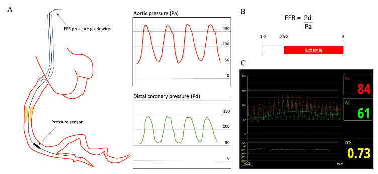
Fractional flow reserve – FFR
The measurement of the coronary flow reserve (FFR) is an invasive modality used to assess the severity of a stenosis. In certain circumstances coronary angiography alone is insufficient to determine if a coronary artery narrowing is severe enough to prevent blood from circulating. The interventional cardiologist can measure the pressure downstream of the stenosis by placing a pressure wire in your diseased coronary artery. The normal ratio is equal to 1, while a ratio 0.8 indicates that the narrowing limits forward blood flow in your coronary artery and therefore requires intervention by angioplasty.

How does FFR takes place?
The use of FFR does not change the course of a coronary angioplasty. It is only a specific device used to find out whether or not it is necessary to intervene in your coronary artery. This exam is painless.
Is it dangerous?
Complications related to the use of FFR are the same as that of the coronary angioplasty.