A coronary angiogram, also referred to as “coronarography”, is the investigation of choice for the visualization of coronary arteries. Your cardiologist has prescribed this exam for the sole purpose of verifying the condition of the arteries supplying your heart. The interventional cardiologist performing this exam can visualize narrowing of the arteries (stenosis) that could lead to shortness of breath, chest pain, myocardial infarction (heart attack) or sudden cardiac death. Visualizing these narrowed coronary arteries enables the selection of the best approach to treat and /or repair your arteries when indicated. If there is any doubt regarding the significance of the narrowing, the cardiologist will use additional specific tools to study the artery (FFR or OCT).
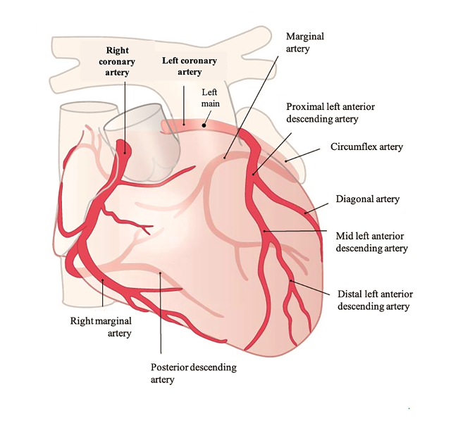
Coronary arteries – what are they?
The coronary artery circulation is comprised of two main coronary arteries: the left coronary artery and the right coronary artery, which supply the heart muscle. They are located at the surface of the heart muscle. The coronary arteries originate at the base of the ascending aorta from openings located behind the leaflets of the aortic valve. They vascularize different myocardial territories through multiple secondary branches and sub-branches.
How does a coronarography takes place and how it is performed?
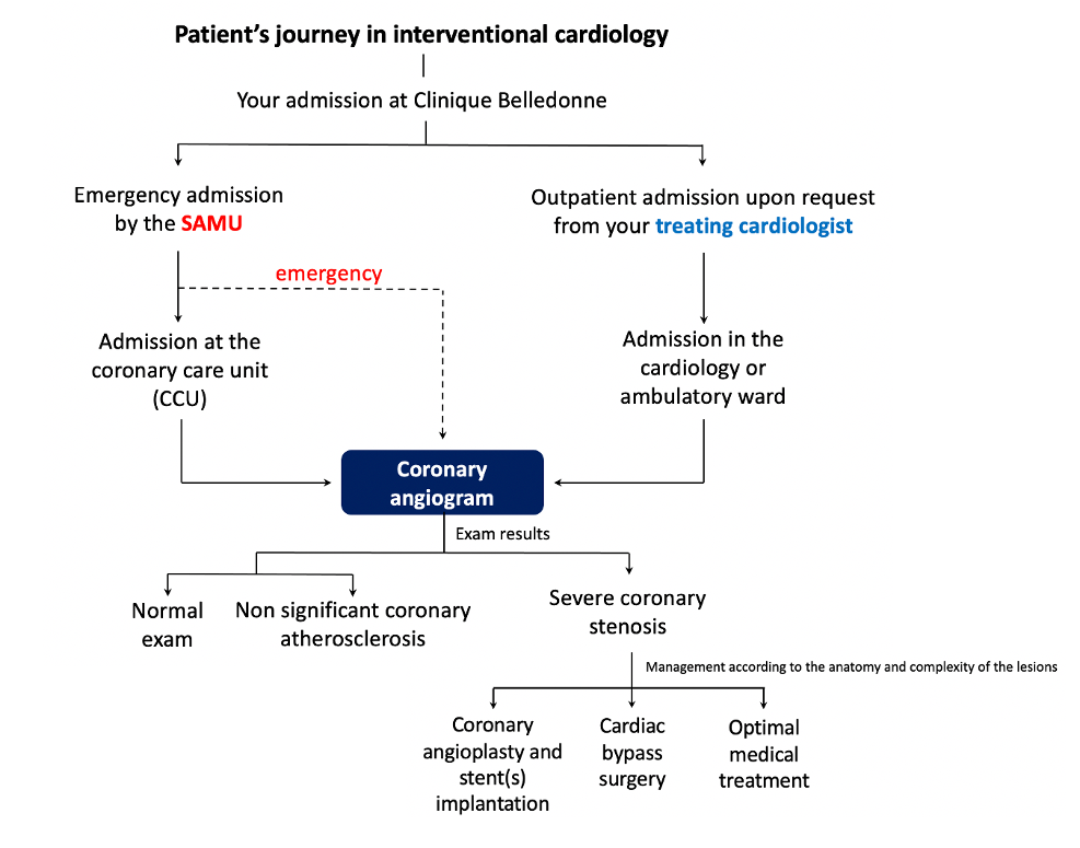
In general, you are hospitalized for 24 hours, with an admission at the Clinique Belledonne the day before the exam around 4pm. Upon a request from your cardiologist, the coronary angiogram can be performed on an outpatient basis, with an admission the morning of your exam and a mid-afternoon medical discharge.
You will be fasting for 6 hours prior to the exam.
In order to avoid infections, a hygiene protocol is necessary, whereby a shower using an antiseptic product and shaving of both wrists (sometimes the groins) is requested.
You will be accompanied from your room to the coronary angiography laboratory by a patient attendant.
Once you are positioned on the catheterization laboratory table, a medication will be administered intravenously by the anesthesiologist that will relax you throughout the entire exam.
The coronary angiography is mostly carried out under local anesthesia through the artery of the wrist (radial artery) or rarely by the groin (femoral artery). The exam is painless for the vast majority of patients. While passing through the radial artery, you may experience a sensation of heat and/or cooling of the forearm and hand that will disappear very quickly.
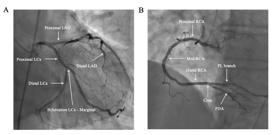
A small diameter catheter is positioned in the coronary arteries under X-ray guidance. It allows to selectively inject an iodinated contrast agent in order to visualize the left and right coronary arteries. A large “C” shaped X-ray camera will rotate around you during the procedure allowing visualization of all your coronary arteries from several different angles.
At the end of the exam, the introducer is removed from the radial artery. A compression dressing is applied on the wrist to avoid bleeding and hematoma formation. The interventional cardiologist will immediately inform you of the results of the diagnostic coronary angiogram. The most suitable treatment will be fully explained to you, in consultation with your treating cardiologist.
You will then return to your room for the rest of the surveillance period and the compression dressing will be removed after 4 hours. In case the exam is performed by the femoral artery, the surveillance period is prolonged.
In general, you are able to return home the same day after a period of surveillance of 6 hours. However, under no circumstances will you be allowed to drive.
Specific conditions
- If you are allergic to contrast media, an antiallergic preparation will be prescribed the day before the exam.
- If you have renal failure, rehydration will be initiated the day before the exam and some of your treatments may be suspended.
- If you have diabetes, your biguanide treatment will be suspended 48 hours prior to the exam.
Is this exam dangerous?
- Despite much technical progress and highly experienced physicians, the coronary angiogram, like any invasive or surgical procedure, carries a risk of incidents or complications.
- Complications are mostly benign and local, such as bleeding or hematoma linked to the arterial puncture site.
- Serious complications are rare (risk of death: 1 per 10,000; risk of stroke: 1 per 1000). Other complications are also possible (less than 1% of cases): myocardial infarction, ventricular rhythm disorder, allergic shock to iodinated contrast media, heart failure decompensation and cardiac perforation.
- All potential complications are fully detailed in the informed consent form for the exam given upon admission and to be signed before the procedure.
In summary
- Hospitalization for 12 to 24 hours.
- Mean duration of the exam: 15-20 minutes.
- The exam is painless for the majority of patients.
- A 6-hour period of monitoring is required following the procedure. In the absence of symptoms and/or bleeding at the puncture site, the patients can return home the same day of the examination (driving is prohibited).
- Strenuous physical effort is discouraged for 72 hours following the procedure to avoid bleeding at the radial and/or femoral puncture site.