
This technique aims to repair the arteries of the legs in order to restore blood circulation in tissues located downstream of a stenosis or occlusion. It consists in positioning a small balloon at the level of the narrowing or the occlusion and to inflate it. By doing so, the atherosclerotic plaque will be crushed against the arterial wall by the balloon, thus allowing the artery to reopen. Most often, the balloon is introduced into the artery by the groin. In certain cases, a stent will be needed at the site of the narrowing or occlusion to maintain the artery patent. The stent is left in place permanently and enables proper blood flow into the leg when balloon angioplasty alone is not effective.
This intervention can improve or relieve your symptoms because it restores oxygen delivery by the arteries to your tissues without requiring surgical intervention.
How does a peripheral angioplasty take place?
A 24 to 48-hour hospitalization is required. The day prior to the intervention, you will be given a blood thinning treatment (antiplatelet agents) to prevent clot formation into the artery.
After being installed on the examination table, a medication will be given by the anesthesiologist to relax you throughout the entire exam.
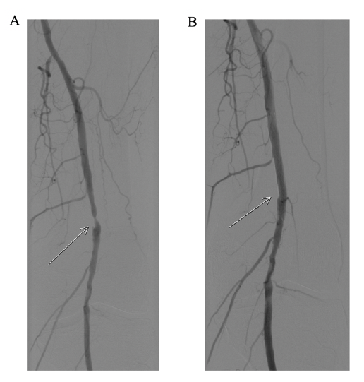
A catheter is introduced most often into the groin and reaches the artery of the leg to be treated. A metal guidewire is then brought into the diseased artery under iodinated contrast media and X-ray guidance. A balloon in inflated to reopen the artery at the site of the obstructive atheroma plaque. Balloon inflation may sometimes be accompanied by pain in the thigh or calf. If the results are not satisfying, the interventional cardiologist may need to put a stent. At the end of the procedure, the guidewire and catheter are removed, and the puncture site is closed with an internal suture. A compressive dressing is positioned on the groin and you will need to lie down for a total of 6 hours.
You will be authorized to leave the clinic the next morning, following the interventional cardiologist’s visit. The doctor will indicate the ideal timing to go back to work and to resume physical activities/sports and/or prescribe a medical leave, if indicated.
You will be given a prescription prior to your discharge from the clinic, which will include a combination of antiplatelet agents (blood thinners) prescribed for several months.
Is it dangerous ?
Complications related to lower limb arteriography are extremely rare.
They are mostly benign and local, such as bleeding or hematoma at the arterial puncture site. Rarely the artery can be injured during the intervention, thus requiring emergent surgical repair or angioplasty. Allergic shock to iodinated contrast media is exceptional. All the potential complications are fully detailed in the informed consent form for the exam given upon admission and to be signed before the procedure.
In summary
- Hospitalization for 24-48 hours.
- The mean duration of the intervention: 30-40 minutes
- The intervention is painless for most patients.
- A 6-hours immobilization period following the intervention is required in order to avoid bleeding at the puncture sites. In absence of symptoms and/or bleeding at the puncture site, the patient can leave the clinic the next morning, following the interventional cardiologist’s visit.
- Rigorous compliance to the medical treatment is essential.
- Strenuous physical effort is discouraged for 72 hours following the procedure to avoid bleeding at the radial and/or femoral puncture site.